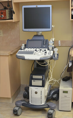
Westbridge Veterinary Hospital has a new, state of the art, Ultrasound machine. Introducing our GE Logiq S 8 ultrasound, taking our ability for diagnostic studies to a higher level of accuracy.
At Westbridge Veterinary Hospital, we offer a very specialized level of ultrasonography skills. Ultrasonography is an incredibly useful diagnostic service for your pet’s health care. An ultrasound is painless and non-invasive. We can “see” into the abdomen of a vomiting cat, the heart of dog with cardiac failure, a swelling or mass to help determine what it is. The skills to do this, however, are very operator dependent. At our hospital two of our veterinarians have shown commitment to advanced study in this modality.
Recently, we have proven our commitment to both animal patient care and excellence through the purchase and upgrading our ultrasound machine to a state of the art GE Logic S8 unit. Through this acquisition, we are excited to offer new non-invasive diagnostic imaging procedures that further benefit our patient’s health and well-being. The goal is to get the right diagnosis for your pet in a gentle and minimally invasive manner that is also cost effective.
Elastography
One such application that we can now provide is Elastography. In many cases, this setting can assist us in differentiating whether a mass within an organ is either malignant or benign. With this revolutionary service we may be able to avoid unnecessary biopsies and the additional stress for your pet. It is also completely painless. Among ultrasonographers we agree this is as close as we can come to having a “histology” button, helping us to provide more information about a tumour. When we sound malignant masses, they will often image as a blue colour with this setting, giving us a value of 4 or 5 on a Tsukuba scale. This is demonstrated in the images below (Figures 1 through 3).
Below are examples of the graphics provided through Elastography.
FIGURE 1: Above, dark blue represents tissue densities that are much firmer than those surrounding it. This can be suggestive of malignant cancer within the liver of a dog.
FIGURE 2: The overall lighter colour in this mass in the neck indicates that it is more likely infection rather than neoplasm (tumour).
FIGURE 3: This firm mass within a kidney is lymphoma. It has a more intense blue colour overall.
High Resolution Imaging
When your pet is ill, finding out what is wrong is of paramount importance. If there is any possibility of cancer, the correct diagnosis is crucial before moving forward with treatment, or other major decisions. The high resolution images below show examples of clear diagnoses.
Below are images of the liver. It should normally be a smooth, homogeneous organ (a similar appearance throughout).
FIGURE 4: Unfortunately is metastatic lymphoma. A cancer that has spread to the liver. It is imaged as a multitude of dark (hypoechoic) focal lesions within the organ. Figure 5 is primary liver cancer and is seen as bright spherical densities within the organ.
FIGURE 5: Primary liver cancer.
Portability
Occasionally we will require ultrasound in an emergency or surgical situation. The compact design of the unit will allow easier access to the operating or treatment room to provide this service. This adds another level of service to our hospital’s diagnostic imaging capabilities.
Cardiology
For our patients with cardiac disease, we have a new cardiac package that can measure the wall motion of the heart chambers to evaluate where there is an abnormality, such as poor heart muscle contractility. This is only offered in a few veterinary hospitals in Canada and we are proud to have it available to our clients. This will prove especially useful for our feline patients in the early detection of cardiac disease. We now also have a new cardiac matrix probe which offer a much higher level of resolution and sensitivity in our cardiovascular studies.
Q Analysis
One such feature for cardiology is called Q Analysis. This calculation tracks the motion of the heart wall along different sections of the organ to look for asymmetry. These fine differences are especially helpful in feline cardiac diagnosis.
Our cardiac patients now have access to a wide variety of cardiac measurements and calculations such as Q Analysis. This helps evaluate cardiac function.
Color Doppler
This setting helps us to view normal and abnormal blood flow within the heart.
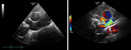
FIGURE 6: The sensitive Matrix cardiac probes on this machine help to detect congenital abnormalities (birth defects) such as the PDA (Patent Ductus Arteriosus) seen in this patient. The yellow-green color represents turbulent blood flow created by the heart defect.
M Mode analysis
This setting helps us to see how well the heart muscle contracts. It views the motion of the heart muscle.
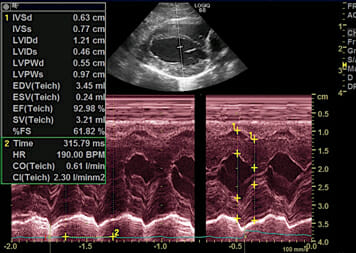
FIGURE 7: This is an example of M mode which studies the heart to help evaluate the severity of heart failure. We can also see improvement with treatment by repeating this after medication has started.
3-D Imaging
Continuing on the long list of capabilities for our ultrasound is the ability to produce 3-D images of structures. This is especially useful for vascular (blood vessel) studies and the search for shunts.
FIGURE 8: This is a 3-D reconstruction of forelimb blood vessels which could look for blood vessel through a tumour for instance.
FIGURE 9: This 3-D image looks at blood vessels within the kidney.
Matrix Transducer Technology
As mentioned in our cardiology section above, these new Matrix transducers offer an enhanced view with higher resolution of small anatomical structures within the body. This allows us to better detect minute changes in specific organs that are diagnostic of certain diseases. This is an important diagnostic feature to have when trying to uncover the root of a complex problem in your pet.
In this high-resolution linear matrix image of the kidney, pus can be seen accumulating in the renal pelvis. This information helped us diagnose a kidney infection.
B Steering
One of the features that we have available on the S8 ultrasound machine is the ability to better image our biopsy instruments as they obtain samples of tissues. This remarkable feature elevates our accuracy when taking tiny biopsy samples for diagnosis. This is critical in the determination of certain conditions. We are proud to have this advantageous feature added to our service.
FIGURE 10: The image above on the left shows a bright image of the biopsy needle advancing into the tissue for a small diagnostic sample. When using the B steer option we can much better visualize the biopsy needle including the shaft and especially the tip.
FIGURE 11: Careful precise placement of a needle is dramatically illustrated with the directional enhancement above.
Logic View Panoramic Imaging
Logic view is another feature that permits us to create continuous images over a large surface area. Below is an image taken over a lower limb. Although the distance measured is over 30 centimeters there is still dramatic clarity. This gives us a panoramic view of a body part. Notice the MR like quality of imaging.
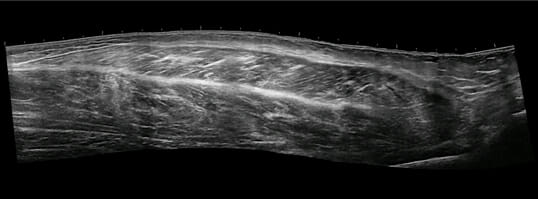
FIGURE 12: Panoramic Imaging of a lower limb.
B Flow
B Flow is a unique feature of GE ultrasound that detects blood flow through the real movement of red blood cells. (A separate feature from the Dopplar).
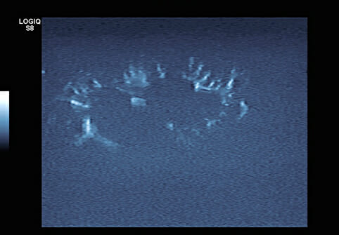
FIGURE 13: An image of flow through the cortex of a kidney using B Flow.
We are happy to tell you more about our Diagnostic Imaging ability at Westbridge Veterinary Hospital. Our commitment is to offer excellence when caring for your pet.