
Pet Diagnostic Imaging – Dental Radiology
Many tooth conditions can be missed without dental X- rays. High resolution digital dental radiographs show the tooth pulp, roots, crown and surrounding bone of each tooth with dramatic clarity. This information helps the veterinarian uncover hidden conditions such as tooth abscesses, cavities and fractures which can cause pain in your pet. Often the owner is not aware of the problem since most animals continue to eat.
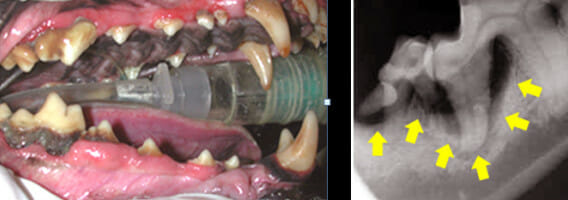
Above left, tartar and plaque covers the base of the tooth and hides potential problems. Above right, on X-ray the arrows outline an area where we can clearly see that all of the jaw bone and associated gum line has receded from the root of the tooth leaving it exposed to disease and pain.
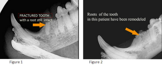
FIGURE 1 (left) and FIGURE 2 (right): In some patients the teeth can be so affected that the crown can actually break away leaving exposed roots (Figure 1 above). There is pain from the tooth disease, the fracture of the tooth, and then the exposed root fragment. There can be remodelling over time (Fig 2), as long as the infection is under control. Digital dental radiographs clearly identify these situations and are essential in helping to plan for the ideal treatment for your pet.
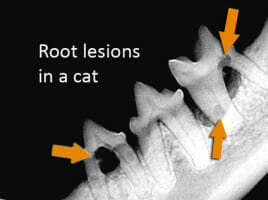
FIGURE 3: These are examples of typical root lesions found in cats. Note that these teeth appeared perfectly normal on physical exam. At our hospital we recommend full mouth dental radiographs for all cats because of this. Most pets that are over 6 years of age have some form of dental disease. It is instinctive for pets not to show pain; therefore a thorough examination is required.
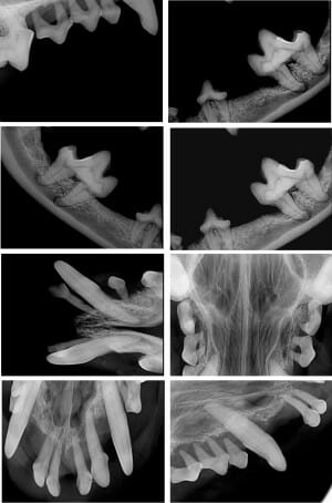
Take a look at this collage of digital images from a dog’s mouth and try and decide which teeth are abnormal.
Disease can affect each complex part of the tooth listed above, either alone or in combination. Without radiographs, we are only seeing and diagnosing disease in the enamel and partially the crown.
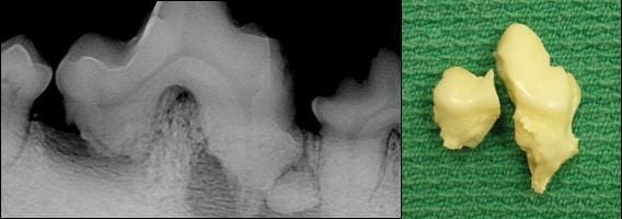
Compare the image on the left, a dental radiograph, with the image on the right, an actual photograph of the same tooth after extraction. Note that the tooth was cut in half to facilitate easier extraction. This is a great example of the fine detail that can be obtained with dental radiography. Take a look at the picture below:
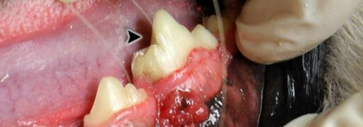
It is a photograph of the same tooth, still intact in the mandible, prior to extraction. Note that visually, above the gum line, there are no abnormalities, and the tooth would otherwise appear to be vital. It is a great display of the importance of dental radiography, as left untreated, this was a very painful tooth.