Pet X-Ray and Ultrasound
X-Rays are not just for broken bones. Radiographs can give us a look at such things as the size of the heart, the pattern of disease in the lungs, intestines, liver, spleen and bladder. Although an x-ray is quick and painless, we may suggest some sedation to assist with the procedure. Dr. Hylands attends internet radiology rounds each Sunday evening with other associate editor veterinarians on the Veterinary Information Network. The information shared during these sessions has been an enormous source of learning new knowledge that is extended to our patient’s care.
How do you use X-ray and radiology services at your hospital?
We now use direct digital radiography, which comes with many benefits to our hospital and our patients! Some of the shining points include:
- Less radiographs due to an increase in diagnostic capability. Digital radiography comes with much higher contrast images. It results in less x-ray exposure to both our patients and staff!
- Less retake x-rays due to imperfect settings. Digital radiography allows us to manipulate the image digitally and is not nearly as sensitive to incorrect settings as plain film. It also reduces the x-ray exposure to our patients.
- Much more environmentally friendly! Plain radiographs require the use of film and x-ray chemicals that must be replaced frequently. The change to digital radiography will result in less chemicals in the environment and less medical waste with old films!
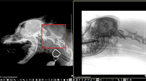
The above side-by-side images of a canine skull were taken with our digital radiography system and have been opened in our software program where it can easily be manipulated. On the left and outlined in red, the magnifying tool is being used, enlarging the area to 1.5x its original size. On the right, the image colours are inverted, allowing for better visualization of some bone structures.
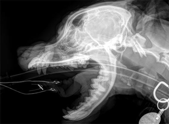
Above is the same skull radiograph zoomed in.
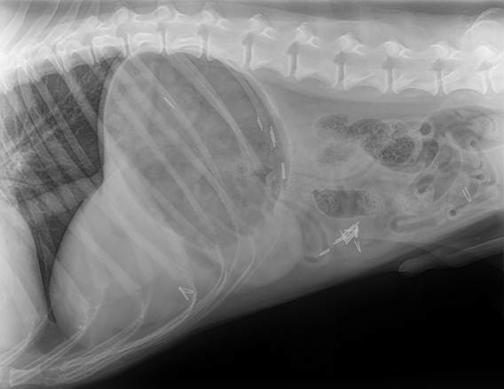
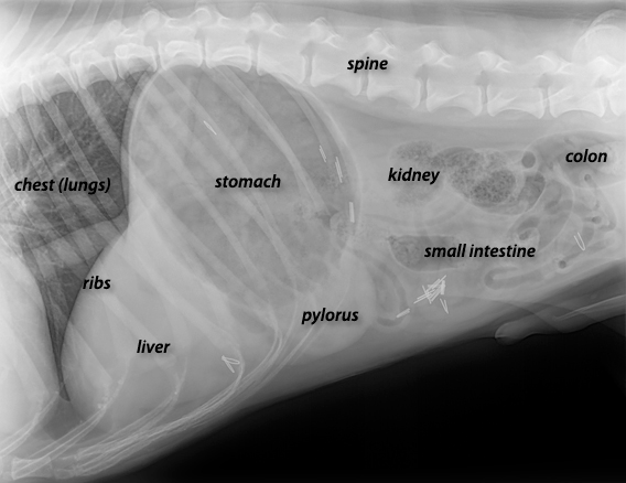
The image above is of a normal abdomen. See if you can match the organs to the image with the lettering (below) when comparing both images.
![]()
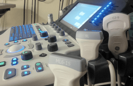
At Westbridge Veterinary Hospital, we offer a high level of ultrasonography skills. Ultrasonography is an incredibly useful diagnostic service for your pet’s health care. An ultrasound is painless and non-invasive. We can “see” into the abdomen of a vomiting cat, the heart of a dog with cardiac failure, swelling or mass to help determine what it is. The skills to do this, however, are very operator dependent. At our hospital, two of our veterinarians have shown a commitment to advanced study in this modality. One of our veterinarians, Dr. Hylands, is also a consultant on the Veterinary Information Network (VIN), where he advises other colleagues on their own individual cases from all over the world. He also teaches instructive seminars all across Canada in the art of ultrasonography.
Recently, we have proven our commitment to both animal patient care and excellence through the purchase and upgrading of our ultrasound machine to state of the art GE Logic S8 unit. Through this acquisition, we are excited to offer new non-invasive diagnostic imaging procedures that further benefit our patient’s health and wellbeing. The goal is to get the right diagnosis for your pet in a gentle and minimally invasive manner that is also cost-effective.
![]()
High-Resolution Imaging
When your pet is ill, finding out what is wrong is of paramount importance. If there is any possibility of cancer, the correct diagnosis is crucial before moving forward with treatment, or other major decisions. The high-resolution images below show examples of clear diagnoses.
Occasionally we will require ultrasound in an emergency or surgical situation. The compact design of the unit will allow easier access to the operating or treatment room to provide this service. It adds another level of service to our hospital’s diagnostic imaging capabilities.
![]()
Cardiology
For our patients with cardiac disease, we have a new cardiac package that can measure the wall motion of the heart chambers to evaluate where there is an abnormality, such as poor heart muscle contractility. This is only offered in a few veterinary hospitals in Canada, and we are proud to have it available to our clients. This will prove especially useful for our feline patients in the early detection of cardiac disease. We now also have a new cardiac matrix probe which offers a much higher level of resolution and sensitivity in our cardiovascular studies.
We are happy to tell you more about our Diagnostic Imaging ability at Westbridge Veterinary Hospital. Our commitment is to offer excellence when caring for your pet.